Advancing Medical Diagnostics: AI-Driven Non-Rigid Image Registration for Precision Healthcare
Explore how ARSA Technology's AI-driven solutions enhance medical image registration, enabling precise diagnosis, treatment planning, and interventions for global healthcare with real-time, accurate alignment.

Medical image registration is a foundational technology in modern healthcare, serving as the invisible backbone for numerous diagnostic and therapeutic procedures. This process involves the spatial alignment of two or more medical images to establish precise anatomical or functional correspondences. From tracking disease progression to guiding intricate surgical interventions, accurate image registration ensures that clinicians can confidently compare, analyze, and act upon critical patient data.
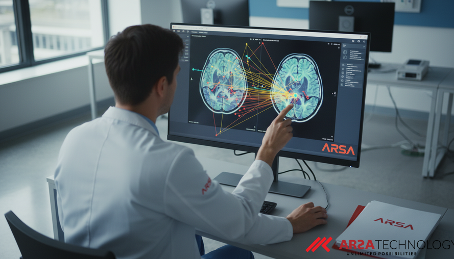
A specialized area within this field is "deformable" or "non-rigid" registration, which accounts for the complex ways tissues and organs can move, stretch, or compress. Unlike rigid transformations that simply shift or rotate an image, non-rigid registration estimates a dense displacement field – essentially, a detailed map of how every point in one image needs to deform to align perfectly with another. This capability is paramount for conditions involving significant anatomical variations or dynamic organ motion, such as lung scans during breathing or tumor growth over time. Despite its importance, achieving this precise alignment, especially with large deformations, while ensuring the resulting image transformation is anatomically logical, remains a significant challenge.
The academic paper, "Attention-Driven Framework for Non-Rigid Medical Image Registration" by Muhammad Zafar Iqbal, Ghazanfar Farooq Siddiqui, Anwar Ul Haq, and Imran Razzak (Source: https://arxiv.org/abs/2602.07088), introduces a novel approach to tackle these challenges. The research proposes an Attention-Driven Framework for Non-Rigid Medical Image Registration (AD-RegNet), leveraging sophisticated AI to guide the alignment process. This framework is a testament to how cutting-edge deep learning, combined with intelligent attention mechanisms, can deliver significant improvements in the accuracy and efficiency of medical image analysis, ultimately enhancing patient care.
The Evolving Landscape of Medical Image Registration
Traditionally, deformable registration relied on complex mathematical optimization tasks. These methods would attempt to maximize the similarity between a fixed reference image and a moving image, while applying mathematical constraints (regularization terms) to ensure the deformation was smooth and anatomically plausible. While effective for certain cases, these conventional techniques often faced hurdles. They were computationally intensive, demanding significant processing power and time, which limited their utility in time-sensitive clinical scenarios like surgical guidance. Furthermore, their sensitivity to parameter settings and their struggle with capturing extremely large deformations meant that consistent, high-quality results were often difficult to achieve.
The advent of deep learning has revolutionized many areas of computer vision, and medical image registration is no exception. Neural networks, with their immense representational power, have proven adept at learning the intricate mappings required to align image pairs. Early deep learning models, such as the seminal VoxelMorph framework, used convolutional neural networks (CNNs) to predict these deformation fields, significantly improving both speed and accuracy. These learning-based methods fall into various categories: supervised (trained on known deformation fields), unsupervised (learning based on image similarity alone), or weakly/semi-supervised (using minimal annotations). While offering substantial advancements, these deep learning approaches still encounter difficulties, particularly in preserving anatomical consistency, handling very large deformations, and intelligently focusing on the specific anatomical structures most relevant to a clinical task.
How Attention Mechanisms Transform Image Alignment
A key innovation in recent AI advancements is the concept of "attention mechanisms." These are powerful tools that allow a neural network to dynamically focus on the most important parts of its input data, much like how a human selectively focuses their attention on specific details while filtering out irrelevant information. In the context of medical image registration, attention mechanisms can be used to pinpoint corresponding anatomical structures within different images and guide the deformation process more effectively.
The AD-RegNet framework, as discussed in the paper, introduces several advanced attention mechanisms to achieve superior registration. One core component is a bidirectional cross-attention module. This module explicitly models the intricate relationships between features extracted from both the moving and fixed images across multiple scales. By establishing these correspondences at different levels of detail, from broad structural outlines to fine-grained textures, the system can achieve more accurate alignments.
Furthermore, the framework incorporates a regional adaptive attention mechanism. This allows the AI to identify and prioritize anatomically relevant structures within the images. For instance, in a thoracic CT scan, the system might focus its attention more intensely on the lungs or heart, knowing these areas are critical for precise analysis. This targeted attention improves registration accuracy precisely where it matters most for clinical evaluation. ARSA Technology, for example, develops advanced AI Video Analytics solutions that utilize similar attention-driven computer vision techniques to focus on critical elements in diverse industrial and commercial settings, enhancing real-time monitoring and operational intelligence.
Beyond Pixels: Ensuring Anatomical Plausibility
Achieving accurate image alignment isn't just about matching pixel values; it's about ensuring that the resulting transformation makes anatomical sense. A deformed image must still represent a biologically plausible structure, without unnatural stretching, tearing, or folding of tissues. The AD-RegNet addresses this through a multi-resolution deformation field synthesis approach. This means that the deformation map is generated by combining information from various resolutions, allowing for robust handling of both global shifts and subtle local deformations, while maintaining a smooth and coherent transformation.
To rigorously validate anatomical plausibility, the researchers employ a suite of evaluation metrics. Beyond standard image similarity measures like Normalized Cross-Correlation (NCC), Mean Squared Error (MSE), and Structural Similarity (SSIM), which quantify how well the images match, they also focus on the Jacobian determinant and Target Registration Error (TRE). The Jacobian determinant is a critical mathematical tool that assesses the local volume changes and smoothness of the deformation. A positive Jacobian determinant across the entire image ensures that no tissue is unnaturally compressed or inverted, preserving topology. TRE, on the other hand, measures the Euclidean distance between corresponding anatomical landmarks after registration, providing a direct assessment of alignment accuracy at clinically significant points. The paper's findings indicate that this attention-guided registration not only improves alignment accuracy but also successfully ensures anatomically plausible deformations, a crucial factor for clinical acceptance.
Real-World Impact and Versatility in Healthcare
The practical implications of such advanced registration frameworks are substantial. By enabling highly accurate and anatomically plausible alignment of medical images, technologies like AD-RegNet can significantly improve various healthcare applications. For disease diagnosis, it facilitates more precise comparisons of scans over time, allowing clinicians to accurately track changes in lesions or organs, crucial for monitoring cancer progression or treatment response. In treatment planning, particularly for radiation therapy or surgery, it ensures that therapeutic interventions are targeted with millimeter-level precision, minimizing damage to healthy tissues. Furthermore, in image-guided interventions, real-time registration can provide surgeons with continuously updated anatomical context during procedures, enhancing safety and efficacy.
The versatility of the proposed method was demonstrated through extensive evaluations on two distinct datasets: DIRLab, which comprises thoracic 4D CT scans (capturing lung movement during respiration), and IXI, which includes brain MRI scans. The ability to perform effectively across different anatomical structures (lungs, brain) and varying imaging modalities (CT, MRI) highlights the framework's robustness and broad applicability in diverse clinical settings. The experimental results confirm that this attention-driven approach achieves performance competitive with state-of-the-art methods, all while maintaining a favorable balance between registration accuracy and computational efficiency. This efficiency is key for integrating such advanced tools into busy clinical workflows, where speed is often as critical as precision. For applications in patient monitoring or pre-screening, ARSA offers the Self-Check Health Kiosk, leveraging AI and IoT for quick, accurate vital sign measurements that can complement advanced image analysis.
Driving the Future of Medical Imaging with AI
The development of attention-driven frameworks for non-rigid medical image registration represents a significant leap forward in AI’s contribution to healthcare. By addressing long-standing challenges in accuracy, anatomical plausibility, and computational efficiency, these technologies are paving the way for more precise diagnostics, safer treatments, and ultimately, better patient outcomes. As AI continues to evolve, its ability to meticulously analyze complex medical data will empower healthcare professionals with unprecedented insights. Companies like ARSA Technology, with their focus on AI and IoT solutions, are at the forefront of translating such academic advancements into practical, deployable tools for global enterprises across various industries, including healthcare.
For organizations looking to integrate cutting-edge AI for enhanced medical imaging, operational efficiency, or comprehensive monitoring, exploring the latest in intelligent solutions is crucial.
Discover how ARSA Technology's AI and IoT solutions can transform your operations. For a free consultation and to learn more about our innovative offerings, please contact ARSA.