Advancing Dental AI: Efficient 3D Tooth Segmentation with Anatomical Precision
Explore a novel AI framework that combines computational efficiency with anatomical accuracy for 3D tooth segmentation from CBCT scans, ideal for clinical deployment.

Introduction: Bridging Efficiency and Accuracy in Dental AI
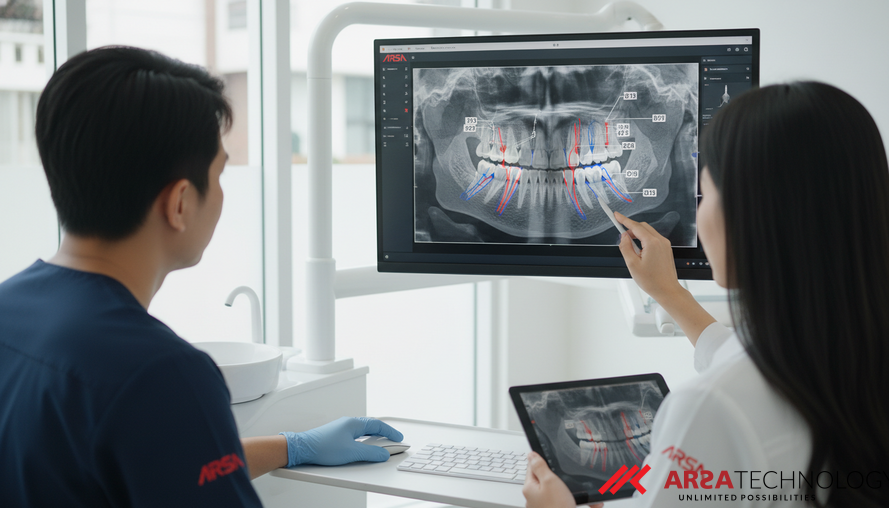
Three-dimensional tooth segmentation, a crucial process in modern dentistry, involves precisely identifying and isolating individual teeth from detailed imaging like Cone Beam Computed Tomography (CBCT) scans. This technology underpins critical applications such as orthodontic planning, implant placement, and even forensic identification. While advanced deep learning models, particularly architectures like nnUNet, have achieved remarkable success in automatically segmenting medical images, their significant computational demands often create a barrier to their widespread adoption in everyday clinical environments. The need for high-performance computing can make these powerful tools impractical where resources are limited.
A common strategy to make deep learning models more efficient is "quantization," a technique that reduces the precision of a model's calculations, thereby decreasing its memory footprint and speeding up processing. However, applying standard quantization to complex tasks like medical image segmentation often comes at a cost: it can degrade the quality of the segmentation, particularly in preserving the intricate anatomical structures and topological relationships between teeth. This compromise between computational efficiency and anatomical fidelity represents a significant hurdle for deploying AI in real-world dental applications. A recent paper, "Topology-Constrained Quantized nnUNet for Efficient and Anatomically Accurate 3D Tooth Segmentation," available on arXiv, addresses this exact challenge.
The Critical Role of 3D Tooth Segmentation
The detailed 3D insights provided by CBCT scans are indispensable for a wide array of dental procedures. For orthodontics, accurate tooth segmentation enables precise treatment planning, allowing practitioners to visualize tooth movement and predict outcomes with greater confidence. In implantology, exact segmentation guides the optimal placement of dental implants, minimizing risks and improving success rates. Beyond these, it assists in forensic identification by providing unique dental signatures. The high stakes involved in these applications mean that any automated segmentation system must deliver not just good but anatomically plausible results. Errors in tooth count, incorrect adjacency, or fragmented structures can lead to misdiagnoses, suboptimal treatment, and potentially severe patient outcomes, highlighting the demand for robust and reliable AI.
The Quantization Dilemma: Speed vs. Anatomical Fidelity
The drive for computational efficiency in AI models is understandable. Quantization, by reducing the numerical precision of a model's weights and activations (e.g., from 32-bit floating-point numbers to 8-bit integers), can dramatically cut down memory usage and accelerate inference. This makes powerful AI models suitable for deployment on less powerful, resource-constrained hardware, like edge devices found in many clinical settings. For instance, ARSA's AI Box Series leverages edge AI to bring powerful analytics directly to where they are needed, reducing cloud dependency and latency.
However, in medical imaging, this efficiency often introduces a critical problem: spatial distortions. Standard quantization methods, while boosting speed, don't explicitly consider the anatomical correctness of the output. This can result in segmented teeth appearing fragmented, merging incorrectly with adjacent teeth, or exhibiting spurious holes—all of which are anatomically incorrect and clinically unacceptable. Current quantization-aware training (QAT) techniques typically optimize for hardware performance, overlooking the vital need for anatomical plausibility, thereby creating a significant trade-off that has limited the clinical utility of otherwise powerful AI tools.
Introducing Topology-Constrained Quantization-Aware Training
To overcome the challenges of traditional quantization, a novel framework has been proposed that integrates specific topological constraints directly into the quantization-aware training (QAT) loop of nnUNet. This innovative approach ensures that the quantized model not only retains its computational efficiency but also produces segmentations that are anatomically valid. Unlike generic anatomical constraints that might apply to various body parts, this method specifically targets the unique topology of teeth. This includes maintaining the correct tooth count, ensuring accurate adjacency relationships between teeth, and eliminating unrealistic "holes" or fragments within tooth structures.
A key advantage of this framework is its ability to achieve anatomical fidelity without requiring any modifications to the underlying nnUNet architecture. This makes it highly compatible with existing implementations and simplifies integration into diverse clinical workflows. By formalizing these dental-specific topological invariants as differentiable loss functions, the model can be trained end-to-end, learning to optimize for accuracy, efficiency, and anatomical correctness simultaneously.
How the Topological Loss Works
The topological loss function is the cornerstone of this innovative approach, designed to complement traditional segmentation accuracy metrics and quantization regularization. It guides the model by penalizing deviations from expected anatomical structures. This is achieved through a combination of several components:
- Connected-Component Analysis: This ensures that each segmented tooth forms a single, continuous object, preventing artificial fragmentation.
- Adjacency Consistency: This component assesses and penalizes incorrect spatial relationships between teeth, ensuring they are separated correctly and don't merge or overlap unnaturally.
- Hole Detection Penalties: These prevent the formation of spurious holes or gaps within the segmented tooth volumes, upholding the integrity of each tooth's internal structure.
The method employs gradient approximations for persistent homology terms, a sophisticated mathematical concept that allows the model to "understand" and maintain structural relationships during training. This joint optimization strategy balances standard cross-entropy loss (for pixel-level accuracy), quantization regularization (for efficiency), and these topological constraints (for anatomical correctness), leading to a highly robust and reliable segmentation solution.
Practical Implications for Clinical Dentistry
The development of topology-constrained quantized AI models holds significant practical implications for the dental industry and beyond. For clinics operating with budget or infrastructure constraints, the ability to deploy highly accurate 3D tooth segmentation AI on less powerful, more affordable hardware (like edge devices) is a game-changer. This translates directly into tangible business outcomes:
- Cost Efficiency: Reduced need for expensive, high-performance computing infrastructure lowers operational costs.
- Faster Diagnostics & Planning: Real-time or near real-time processing of CBCT scans allows dentists to make quicker, more informed decisions for orthodontic treatment, implantology, and other procedures, enhancing patient throughput.
- Improved Treatment Quality: Anatomically accurate segmentations lead to more precise treatment plans, reducing errors and improving patient outcomes.
- Enhanced Data Privacy: On-premise processing options, like those facilitated by edge AI systems, mean sensitive patient data can remain within the clinic's local network, addressing crucial privacy and compliance concerns (e.g., GDPR/HIPAA), a core offering from companies that have been experienced since 2018 in developing robust AI solutions.
- Scalability: The ability to scale deployments across multiple clinics without a massive infrastructure overhaul makes advanced AI accessible to a broader range of dental practices.
Key Findings and Future Outlook
Experiments conducted using this topology-constrained quantization-aware training framework have shown compelling results. Compared to conventional quantized models, the proposed approach significantly reduces topological errors, achieving clinically plausible segmentations on real dental CBCT scans. Crucially, this enhanced anatomical accuracy is delivered while retaining the hardware efficiency inherent to integer-only inference, making these models highly suitable for deployment in resource-constrained clinical environments. The method also maintains competitive Dice scores, a common metric for segmentation accuracy, indicating that the topological improvements do not come at the expense of overall segmentation quality.
This work marks a significant step forward in bridging the gap between computational efficiency and anatomical precision in medical image segmentation. While the immediate focus is on dental applications, the principles of integrating topology-aware losses into quantized models can be extended to other areas of medical imaging, where preserving complex anatomical structures is paramount. Future research may explore adapting this methodology to other types of medical scans or for real-time monitoring applications, continuing to push the boundaries of practical AI in healthcare.
Conclusion: The Future of Precise and Practical Dental AI
The convergence of AI efficiency and anatomical precision is no longer a distant goal but a present reality, exemplified by innovations in topology-constrained neural networks. By enabling deep learning models to perform complex 3D tooth segmentation with high accuracy and anatomical fidelity on resource-efficient hardware, this technology is poised to transform dental diagnostics and treatment planning. The implications are profound, promising more accessible, cost-effective, and reliable AI tools that empower dental professionals and ultimately improve patient care.
To explore how advanced AI and IoT solutions can transform your operations with practical, proven, and profitable deployments, we invite you to contact ARSA for a free consultation.