AI-Powered Chest X-ray Analysis: Predicting ICU Escalation in COVID-19 Patients
Explore how advanced AI and kernel-based learning (GLIMARK) analyze chest X-rays to predict ICU escalation in COVID-19 patients, optimizing resource allocation and enhancing healthcare efficiency.

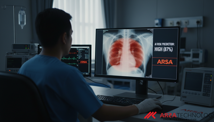
In the rapidly evolving landscape of healthcare, Artificial Intelligence (AI) and machine learning are transforming diagnostic processes and predictive analytics, especially in critical care scenarios. A groundbreaking study introduces an advanced kernel-based learning approach, known as Generalized Linear Models with Integrated Multiple Additive Regression with Kernels (GLIMARK), to interpret chest X-ray (CXR) images. This innovative method aims to predict the likelihood of COVID-19 patients escalating to Intensive Care Units (ICU), offering crucial insights for resource allocation and early intervention strategies, as detailed in the academic paper, "Kernel-Based Learning of Chest X-ray Images for Predicting ICU Escalation among COVID-19 Patients."
The Critical Need for Predictive Analytics in Healthcare
Medical imaging plays a pivotal role in disease diagnosis, prognosis, and outcome prediction. During the COVID-19 pandemic, the ability to accurately forecast patient deterioration, particularly the need for ICU admission, became paramount due to severe resource shortages. Traditional methods of evaluation, while useful, often rely on subjective scoring systems or lack the data-driven depth required for highly accurate predictions. Chest X-rays offer distinct advantages over more complex imaging like CT scans: they are faster, portable, and more accessible, making them ideal for rapid decision-making in high-pressure medical environments.
However, medical images like CXRs have been underexplored for predicting ICU escalation. Existing studies have shown correlations between CXR scores and CT scans with ICU admission, but a more robust, data-driven approach was needed to unlock their full potential for predictive modeling. The challenge lies in extracting comprehensive, clinically meaningful information from these images and integrating it with other patient data to build reliable predictive models.
Overcoming Limitations with Advanced Kernel-Based Learning
Traditional machine learning methods, especially single kernel approaches, often struggle to capture the full complexity and heterogeneity of real-world medical data. These methods might rely on a single type of mathematical function (a "kernel") to find patterns, which can be insufficient when dealing with diverse patient information and intricate image features. This often leads to models that may miss subtle yet crucial indicators.
Multiple Kernel Learning (MKL) emerged as a solution, allowing the construction of composite kernels by combining simpler ones. This enables the integration of information from heterogeneous sources, providing a more comprehensive understanding of the data. Despite these advancements, most conventional MKL techniques were primarily designed for continuous outcomes, limiting their applicability to diverse data types, such as binary outcomes like "ICU escalation" (yes/no). Additionally, some existing MKL methods prioritize computational efficiency or dimension reduction, often at the expense of model interpretability, which is vital in clinical settings.
Introducing GLIMARK: A Generalizable and Interpretable Approach
To address these critical limitations, the study proposes an innovative extension to MKL called Generalized Linear Models with Integrated Multiple Additive Regression with Kernels, or GLIMARK. This new method introduces a likelihood-based loss function within the Generalized Linear Model (GLM) framework. This allows GLIMARK to accommodate a broader range of outcome variables beyond just continuous data, including binary (yes/no), Poisson (count data), and Gaussian outcomes, significantly expanding its utility for diverse medical applications.
A key innovation in GLIMARK is its hierarchical framework for modeling radiomics features extracted from CXR images. Radiomics involves quantifying detailed characteristics from medical images—features like texture, shape, and intensity—that are often too subtle for the human eye to consistently discern. GLIMARK explores these feature patterns at multiple scales, striking a balance between model complexity and its capacity to learn intricate patterns. Unlike approaches that can obscure the meaning of original features, GLIMARK maintains interpretability by focusing on specific feature-kernel combinations, thereby allowing clinicians to understand which image characteristics are driving predictions.
Practical Application and Clinical Significance
The GLIMARK method was rigorously applied to a real-world COVID-19 chest X-ray dataset from Michigan Medicine, an extensive multimodal dataset including electronic health records and nearly two million CXR images. The primary goal was to predict the binary outcome of ICU escalation. The results demonstrated GLIMARK's effectiveness in accurately predicting which patients would require intensive care, thereby enabling more informed and timely decision-making.
A significant finding was the identification of clinically meaningful features that traditional models often miss. Specifically, the study highlighted the importance of Neighboring Gray Tone Difference Matrix (NGTDM) and Shape2D features as top-ranked indicators for ICU escalation. NGTDM features capture image texture and contrast, reflecting changes in tissue density that could indicate disease progression, while Shape2D features describe the morphology of lung lesions. These insights were not detected by conventional regression or random forest models, underscoring GLIMARK's superior ability to uncover complex patterns. Furthermore, the method can identify "representer" patients, whose CXR images exhibit key visual features strongly associated with predicted outcomes. These representers serve as invaluable benchmarks for future radiological analysis and outcome prediction.
Such advanced analytics can be instrumental for healthcare providers globally. For instance, solutions like ARSA AI Video Analytics or bespoke AI model development can be tailored to similar predictive tasks, integrating image analysis with clinical data to provide real-time support for medical staff.
Looking Ahead: The Future of AI in Medical Imaging
The development of GLIMARK represents a significant step forward in leveraging AI for critical healthcare decisions. By transforming medical images into actionable intelligence, it offers a powerful tool for optimizing patient care, improving resource allocation, and potentially saving lives. The method's strengths lie in its generalizability, interpretability, and ability to process diverse data types, making it applicable beyond COVID-19 to other conditions where early prognosis from medical imaging is crucial.
Future research could explore integrating GLIMARK with other advanced imaging modalities, such as lateral view CXRs, and incorporating next-generation AI architectures like Vision Transformers. These advancements could further enhance the accuracy and scope of predictive models, cementing AI's role as an indispensable partner in modern medicine. This kind of advanced analytical capability highlights the potential for comprehensive AI-driven health monitoring systems, much like the insights offered by ARSA's Self-Check Health Kiosk for initial patient screening and data collection.
As AI technologies continue to mature, the focus remains on developing solutions that are not only powerful but also practical, interpretable, and seamlessly integrable into existing healthcare infrastructures. Providers like ARSA Technology, which has been experienced since 2018 in delivering AI & IoT solutions, are at the forefront of this transformation, ready to partner with medical institutions to deploy these life-changing innovations.
To explore how AI and IoT can transform your healthcare operations with measurable impact and efficiency, we invite you to discover ARSA Technology's solutions.
Contact ARSA today for a free consultation.
Source: Kernel-Based Learning of Chest X-ray Images for Predicting ICU Escalation among COVID-19 Patients