AI Revolutionizes Cardiac CT: Enhancing Diagnosis by Eliminating Calcium Artifacts
Discover how Dense-MAE, an AI-powered solution, enhances Coronary CT Angiography (CCTA) by accurately removing calcium blooming artifacts, enabling clearer diagnostics and improving patient outcomes.

The Critical Challenge of Coronary Artery Imaging
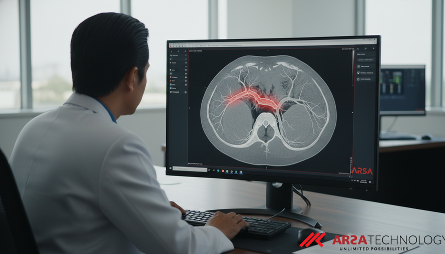
Coronary Artery Disease (CAD) remains a leading cause of mortality worldwide, making accurate and timely diagnosis crucial. Coronary CT Angiography (CCTA) has emerged as a powerful non-invasive tool for visualizing the coronary arteries and detecting blockages. However, a significant hurdle in CCTA interpretation is the presence of "calcium blooming artifacts." These artifacts, caused by dense calcifications in the arteries, often appear larger and blurrier than they truly are, obscuring the actual vessel lumen and complicating diagnosis. This phenomenon can lead to inaccurate assessments, potentially resulting in unnecessary further invasive tests, increased patient anxiety, and higher healthcare costs.
The diagnostic paradox of CCTA is that while it is excellent for detecting calcification, these very calcifications can hinder the clear visualization of the vessel pathways themselves. Addressing this blooming artifact is paramount to unlocking the full potential of CCTA as a precise diagnostic instrument.
Understanding the "Blooming" Problem in CT Scans
Calcium blooming artifacts are a common issue in computed tomography (CT) scans, particularly in cardiovascular imaging. When X-ray beams pass through highly dense materials like calcified plaques in the coronary arteries, the resulting image can show the calcium deposits as artificially enlarged and poorly defined. This "blooming" effect makes it challenging for radiologists to accurately gauge the degree of narrowing (stenosis) in the vessel. Beyond blooming, other artifacts like beam hardening and photon starvation also contribute to reduced image quality, further impacting diagnostic confidence.
Traditional methods have attempted to mitigate these issues, ranging from specialized image reconstruction algorithms like Iterative Reconstruction (IR) and Filtered Back Projection (FBP) to advanced CT techniques such as CT Zoom and Photon-Counting CT (PCCT). While these methods offer some improvements, they often fall short of completely eliminating the pervasive blooming artifacts, especially in patients with extensive calcification. The inherent physical limitations of X-ray imaging mean that a new approach is needed to overcome these persistent challenges.
AI-Powered Solutions for Enhanced Medical Imaging
The advent of Artificial Intelligence (AI), particularly deep learning, has opened new frontiers in medical image processing. Deep Convolutional Neural Networks (DCNNs) and Generative Adversarial Networks (GANs) have shown promise in various image enhancement tasks, including calcium removal. Early applications of DCNNs, such as those employing a Dense-Unet architecture, have demonstrated the ability to reduce artifacts by learning from existing image data. However, these methods sometimes struggle with the "hallucination" problem, where the AI might generate details that aren't truly present, or perform sub-optimally when training data is limited.
The challenge lies in developing AI models that can accurately remove artifacts without sacrificing real anatomical details or introducing false information. This requires intelligent algorithms that understand anatomical context and can learn effectively even from smaller datasets—a common scenario in specialized medical imaging. Solutions like ARSA AI Video Analytics leverage advanced AI vision to address complex visual data challenges, offering a blueprint for similar innovations in healthcare.
Introducing Dense-MAE: A Breakthrough in Medical Image Reconstruction
A significant innovation in this field is the introduction of Dense-MAE, a novel AI model specifically designed for coronary calcium removal in CCTA. Dense-MAE combines the strengths of Masked Autoencoders (MAE) with a Dense-Unet architecture, tailored for 3D volumetric data. The core idea of a Masked Autoencoder is to train an AI by showing it incomplete images (where parts are "masked" or hidden) and asking it to reconstruct the missing information. This forces the AI to learn deep contextual understanding and robust feature representations.
Dense-MAE enhances this concept with several key innovations:
- Vessel-Aware Masking Strategy: Unlike generic masking, this strategy intelligently masks areas of the CT scan while prioritizing the preservation of critical vessel structures, ensuring that essential diagnostic information is not inadvertently removed or distorted.
- Dense-Unet Architecture: This highly interconnected deep learning network is specifically chosen for its superior ability to learn intricate features and reconstruct images with high fidelity. Its "dense blocks" and "3D skip connections" enable efficient information flow and powerful representation learning from 3D volumetric data.
- Anatomical Consistency Constraints: During reconstruction, the model is guided by constraints that ensure the generated images remain medically plausible, effectively mitigating the "hallucination" problem seen in simpler MAE models.
- Self-Supervised Learning (SSL): This allows the model to learn effectively from unlabeled data, reducing the reliance on extensive, manually annotated datasets which are often scarce and expensive to produce in medical imaging.
This combination allows Dense-MAE to achieve robust performance even in "few-shot" learning scenarios, meaning it requires significantly less labeled data for effective training compared to other supervised learning models.
How Dense-MAE Works: A Glimpse into the Technology
The Dense-MAE system processes 3D patches of CT images. In its self-supervised training phase, approximately 60% of an input image patch is intentionally masked in a vessel-aware manner. The Dense-Unet then acts as both an encoder, learning from the visible portions, and a decoder, reconstructing the masked parts. The model's performance is measured using a loss function like Mean Squared Error (MSE), comparing its reconstructed output against the original, unmasked image. This iterative process allows the AI to develop a sophisticated understanding of coronary anatomy and the characteristics of calcium artifacts.
Crucially, the Dense-Unet's design, featuring dense blocks and 3D skip connections, allows it to learn hierarchical features and contextual information across the entire 3D volume. This capability is vital for accurately distinguishing between genuine vessel structures and artifactual blooming. The use of edge computing principles, often facilitated by devices like the AI Box Series, means that this complex processing can occur locally, ensuring patient data privacy while delivering real-time insights directly at the point of care.
Significant Results and Real-World Impact
The Dense-MAE model demonstrated remarkable improvements in performance compared to previous methods. In experiments using 60 CTA volumes, Dense-MAE achieved a PSNR (Peak Signal-to-Noise Ratio) of 32.45, significantly outperforming a standard Dense-Unet (29.88) and a 3D-MAE based on Vision Transformers (28.12). This represents a 2.57 dB improvement in image quality over the traditional Dense-Unet. Furthermore, the Dice Score (DSC), a measure of segmentation accuracy, increased by 4%, reaching 0.905, indicating a much better delineation of relevant anatomical structures after artifact removal.
These quantitative improvements translate into tangible benefits in clinical practice. Clearer CCTA images allow cardiologists to more accurately assess the severity of coronary stenosis, leading to more confident diagnoses of CAD. The ability to achieve robust performance with limited training data, as demonstrated by strong results even with only 25% of the original training set, makes Dense-MAE a practical solution for healthcare providers who may not have vast, annotated datasets. This technology can significantly reduce the risk of misdiagnosis, optimize patient care pathways, and enhance the overall efficiency of cardiac imaging services. Businesses can integrate such advanced AI capabilities through flexible solutions like the ARSA AI API, enabling seamless adoption into existing medical systems.
The Future of AI in Healthcare Diagnostics
The advancements seen with Dense-MAE underscore the transformative potential of AI in healthcare, particularly in improving diagnostic accuracy for critical conditions like CAD. By tackling complex imaging challenges such as calcium blooming artifacts, AI empowers medical professionals with clearer, more reliable data, facilitating better clinical decisions and ultimately leading to improved patient outcomes. This is a crucial step towards precision medicine, where diagnostic tools are finely tuned to provide the most accurate and personalized insights possible.
Beyond coronary calcium removal, the principles of self-supervised learning and anatomically aware AI models can be extended to address other persistent challenges in medical imaging and diagnostics. ARSA Technology, with a team experienced since 2018 in AI and IoT solutions, is committed to pioneering these innovations. From enhancing imaging for various diseases to developing smart diagnostic tools, the future holds immense promise for AI-driven healthcare. This technology can be adapted not only for high-end hospital systems but also for broader public health initiatives, akin to how solutions like the Self-Check Health Kiosk make basic health monitoring accessible to a wider community.
Ready to explore how advanced AI can transform your healthcare operations and diagnostic capabilities? Discover ARSA Technology's innovative solutions and contact ARSA for a free consultation.