Beyond Simulation: AI Diffusion Models Create Hyper-Realistic PET Images for Medical Advancement
Explore how a novel AI diffusion model, PAD, synthesizes highly realistic, heterogeneous PET images from simple organ maps. This breakthrough revolutionizes medical research, AI training, and virtual trials by overcoming traditional simulation limits. Learn about its precision and real-world applica

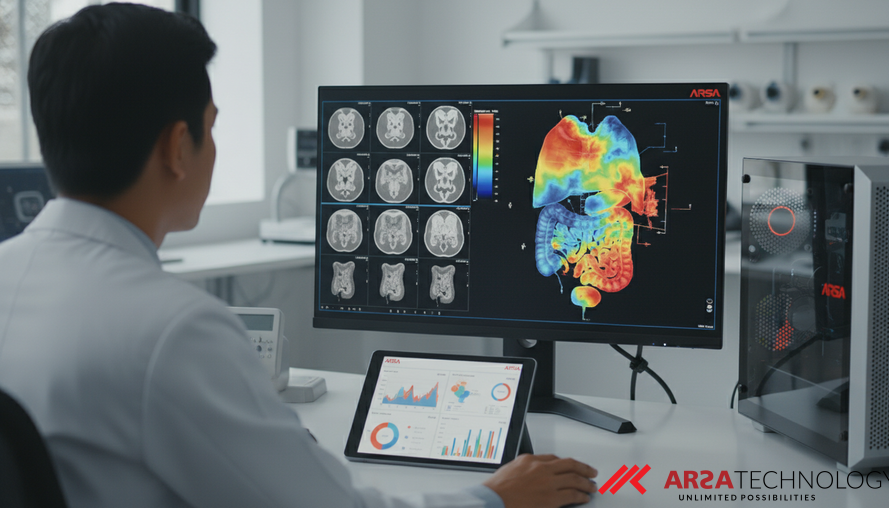
In the rapidly evolving landscape of medical technology, Positron Emission Tomography (PET) imaging stands as a crucial diagnostic tool. PET scans offer a unique window into the body's metabolic activity, providing vital information for detecting diseases like cancer, neurological disorders, and cardiovascular conditions. However, the development, optimization, and validation of new PET imaging techniques often face a significant hurdle: the scarcity of high-quality, diverse, and representative clinical imaging data. This limitation has historically led researchers to rely on synthetic PET images, but traditional generation methods have presented their own set of challenges.
The Critical Need for Realistic Synthetic PET Images
Synthetic PET images are invaluable for a multitude of applications within quantitative imaging workflows, enabling scalable virtual imaging trials, and providing essential data for training sophisticated deep learning models. Traditionally, these synthetic images are created using physics-based simulations. Methods often involve digital anthropomorphic phantoms, such as the 4D extended cardiac-torso phantom (XCAT), combined with simulation tools like Geant4 Application for Tomographic Emission (GATE). While these tools offer reproducibility and precise control over scanner settings, they come with significant drawbacks.
Running a GATE simulation for a single patient can take many hours, making large-scale data generation impractical. More critically, these simulations frequently fail to capture the subtle yet crucial uptake heterogeneity and realistic visual appearance observed in actual clinical PET images. Furthermore, digital phantom designs often lack the flexibility to customize patient anatomy, leading to synthetic images that don't faithfully represent real-world anatomical variations. These limitations make traditional simulated images less suitable for advanced applications like medical image segmentation and harmonization, which are sensitive to fine image details. The growing demand for robust deep learning models in medical image analysis further underscores the need for vast, diverse, and realistic datasets that traditional simulations cannot efficiently provide.
Evolution of AI in Image Synthesis
The past decade has seen remarkable advancements in deep learning techniques for image synthesis, each addressing previous limitations. Early generative models like Variational Autoencoders (VAEs) were capable of scalable image generation but often produced overly smooth images lacking intricate structural details. Generative Adversarial Networks (GANs) emerged as a significant improvement, introducing adversarial training to produce sharper and more realistic images. GANs have found broad applications in medical imaging, including modality translation and image denoising. However, GANs can be notoriously difficult to train, often suffering from instability and a phenomenon known as mode collapse, which limits the diversity of generated outputs.
Recognizing these challenges, Denoising Diffusion Probabilistic Models (DDPMs) have risen as a powerful alternative. Unlike GANs, diffusion models operate by gradually adding noise to data through a forward Markov process. A neural network is then trained to iteratively reverse this process, effectively "denoising" random data step-by-step to generate complex and realistic samples. This approach offers more stable optimization and has proven to outperform GANs in terms of both image fidelity and sample diversity in natural and medical image synthesis. This robust performance has fueled the increasing adoption of diffusion models in various PET imaging applications, paving the way for more sophisticated and reliable synthetic data generation.
Introducing the Pretrained Domain-Adapted Diffusion (PAD) Model
A recent study introduced a novel Pretrained Anatomy-conditioned Diffusion (PAD) model designed to generate realistic clinical PET images from simplified inputs known as uniform organ activity maps. This innovative model leverages the strengths of diffusion models while addressing the unique complexities of medical image data. The PAD model strategically combines a natural-image pretrained text-to-image decoder with a specialized upstream conditioning encoder and a downstream PET-domain adapter. This architecture allows the model to benefit from the extensive knowledge gained from training on vast natural image datasets, while simultaneously adapting that knowledge to the specific nuances and characteristics of medical PET images. For a deeper understanding of how specialized AI models can be integrated into custom solutions, explore ARSA's custom AI solutions.
Generating high-resolution medical images with deep learning requires substantial computational resources and extensive datasets. To overcome these limitations, the PAD model employs a two-phase training strategy. The first phase focuses on learning coarse uptake distributions, essentially mapping out the general patterns of tracer activity within organs. The second phase then refines these initial distributions, adding local image details and realistic heterogeneity. This coarse-to-fine approach is akin to an artist first sketching a broad outline and then meticulously adding fine textures and shading. Furthermore, to enhance training stability and data efficiency, the model leverages a text-to-image model initially trained on natural images. This pretraining provides a rich semantic foundation, stabilizing the learning process when adapting to the specialized PET domain.
From Simplified Inputs to Detailed Outputs
The elegance of the PAD model lies in its ability to transform relatively simple inputs into remarkably complex and detailed outputs. The study utilized a dataset of 513 FDG-PET/CT images to extract patient anatomy. From CT-based segmentations of 128 anatomical structures, uniform organ activity maps were generated by assigning a mean uptake value to each organ, based on its activity in the paired clinical PET image. These uniform maps, essentially simplified representations of organ activity, served as the conditioning inputs for the PAD model, with the corresponding clinical PET images acting as the target outputs.
A crucial step in preparing medical imaging data for deep learning is normalization, which accounts for the wide variability in tracer uptake among patients and the extensive intensity distribution within each PET image. The study implemented a two-step normalization process: first, converting images to Standardized Uptake Value (SUV) to correct for patient body weight and injected dose, reducing inter-patient variability. Second, an inverse hyperbolic sine (arcsinh) transformation was applied to handle the high dynamic range typical of PET images, compressing extremely high intensities while preserving details in regions of interest. This meticulous data preparation improves numerical stability and ensures consistent gradient behavior during the diffusion-based generation process, enabling the AI to learn more effectively.
Validating the Accuracy and Realism of PAD
The credibility of any AI model in medical applications hinges on rigorous validation. The study meticulously evaluated the PAD-generated images across five critical aspects: quantitative accuracy, noise assessment, radiomic analysis, tumor segmentation performance, and a human observer study. These evaluations collectively demonstrated the model's capacity to produce clinically relevant and realistic synthetic images, supporting advanced applications like AI Video Analytics for detailed analysis.
Quantitative accuracy was high, with concordance correlation coefficients exceeding 0.92 between the organ mean SUVs of the synthesized images and their assigned activity values, indicating precise replication of uptake levels. The synthesized images exhibited noise levels and texture characteristics remarkably similar to the target clinical PET images, an important aspect for visual realism and diagnostic utility. Radiomic analysis further confirmed that the generated images captured complex textural features consistently. In a task-based validation, the PAD-generated images produced comparable tumor segmentation performance to actual PET images, demonstrating their utility for downstream clinical applications. Perhaps the most compelling validation came from a two-alternative forced-choice human observer study, where four expert readers achieved approximately 50% accuracy in distinguishing between synthesized and target images. This near-chance performance strongly suggests that the PAD-generated images were visually indistinguishable from real PET scans, a true benchmark for generative AI in medicine.
Broader Implications for Medical Imaging and Beyond
The development of the PAD model represents a significant leap forward in medical imaging technology. By efficiently generating highly realistic, heterogeneous PET images, it addresses critical gaps in data availability for deep learning model training, accelerating the development and validation of new diagnostic tools. This capability is particularly impactful for applications such as creating scalable virtual imaging trials, where new drugs or treatment protocols can be simulated and tested on diverse virtual patient populations without the logistical and ethical complexities of real-world trials.
Furthermore, the model's compatibility with XCAT-derived activity maps bridges the gap between traditional phantom-based simulations and cutting-edge AI, offering a flexible framework for future research. This innovation not only empowers data augmentation strategies for AI-driven medical analysis but also provides a powerful tool for optimizing scanner designs and refining imaging and reconstruction protocols. Companies like ARSA, with extensive experience since 2018 in AI and IoT solutions across various industries, understand the practical deployment realities of such advanced AI. Solutions akin to the PAD model, capable of running on edge devices through products like the AI Box Series, ensure privacy-by-design and low latency processing for sensitive medical data. The ability to generate such high-fidelity synthetic data means faster innovation, reduced costs, and ultimately, improved patient outcomes in diagnostics and treatment planning (Suya Li et al., 2024, "Generation of Heterogeneous PET Images from Uniform Organ Activity Maps Using a Pretrained Domain-Adapted Diffusion Model").
Ready to explore how advanced AI and IoT solutions can transform your operations? Our team is dedicated to engineering intelligence into mission-critical applications across various sectors.
Discover how ARSA Technology can provide tailored AI and IoT solutions for your specific enterprise needs. We invite you to contact ARSA for a free consultation.