Revolutionizing Female Pelvic Health: AI-Powered Anomaly Detection for Real-Time MRI
Discover how Unsupervised AI is transforming diagnosis of female pelvic diseases in real-time MRI, addressing anatomical variability and enabling earlier detection without extensive labeled data.

The Unseen Challenge: Diagnosing Female Pelvic Diseases
Pelvic diseases in women of reproductive age pose a significant global health burden, affecting approximately 40% of this demographic and contributing substantially to worldwide disease. Conditions such as uterine myomas, adenomyosis, endometriosis, ovarian tumors, and various cancers present unique diagnostic challenges. A major hurdle is the inherent anatomical variability of the female pelvis, including differing uterus positions, which complicates the interpretation of Magnetic Resonance Imaging (MRI) scans. This complexity often leads to delayed diagnoses, misclassifications, and prolonged suffering for patients. For instance, endometriosis, affecting about 10% of women, frequently faces an average diagnostic delay of eight years, with nearly 75% of patients initially misdiagnosed.
While ultrasound is a common initial diagnostic tool, its interpretability can be subjective and prone to acoustic shadowing. MRI, however, plays a crucial role in more intricate cases due to its superior spatial resolution, excellent soft tissue contrast, and reduced operator dependence. Despite these advantages, the lack of standardized protocols and the often-oblique positioning of the uterus in pelvic MRIs further complicate diagnosis, which heavily relies on the clinician's expertise and the quality of images captured during the scan. The integration of artificial intelligence (AI) has shown promise in medical imaging, particularly in more invariant areas like brain or chest scans. However, applying these methods to the highly variable anatomy of the abdomen and pelvis introduces unique challenges for AI generalization.
Revolutionizing Diagnosis: The Promise of Unsupervised Anomaly Detection
Traditional AI approaches in medical imaging, which often rely on supervised learning, struggle with the vast and varied manifestations of pelvic diseases. It is simply not feasible to collect and label abnormal data for every possible pathology, leading to models that are disease-specific and lack broad applicability. Furthermore, the clinical reality of inter-observer variability in expert annotations for pelvic MRI images makes creating consistent, labeled datasets for supervised deep learning a complex and often subjective task. This highlights a critical need for alternative AI strategies that can adapt to the unpredictable nature of disease presentation.
Unsupervised Anomaly Detection (UAD) emerges as a highly promising solution to these limitations. UAD models are trained exclusively on "normal" or healthy data, learning the expected patterns and structures without needing any examples of disease. This mirrors how human experts often identify anomalies: by first understanding what healthy anatomy looks like and then recognizing deviations from that norm. By training deep learning models solely on healthy data, UAD can highlight regions in a new scan that deviate significantly from the learned healthy structure, indicating potential pathology without requiring pre-labeled abnormal cases. This approach offers enhanced generalizability and adaptability, making it suitable for complex anatomical regions like the female pelvis (Knupfer et al., 2026, https://arxiv.org/abs/2602.06179).
Behind the AI: How the System Identifies Anomalies
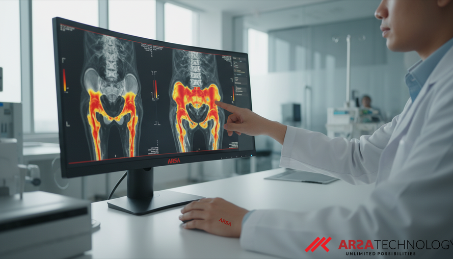
The framework for disease- and parameter-agnostic unsupervised anomaly detection in pelvic MRI utilizes a sophisticated AI architecture known as a residual variational autoencoder. A Variational Autoencoder (VAE) is a type of neural network that learns to encode input data into a compressed representation and then reconstruct it. The "residual" component adds pathways that allow information to bypass certain layers, improving the network's ability to learn complex patterns and reconstruct images more accurately. For this system, the VAE was specifically trained on healthy sagittal T2-weighted (T2w) scans. Sagittal scans provide a side-view of the pelvis, and T2w MRI sequences are optimized to highlight fluid-filled structures and various tissues, making them ideal for pelvic imaging.
To ensure robustness and account for the considerable diversity of acquisition parameters across different imaging protocols, the model was trained on a dataset of 294 healthy scans. This real data was further augmented with diffusion-generated synthetic data. Diffusion models are advanced generative AI models capable of creating highly realistic, AI-generated mock data, which helps improve the VAE’s understanding of normal anatomical variations and enhances its ability to generalize. During the inference phase – when the system analyzes a new, unseen MRI scan – the VAE attempts to reconstruct the input. Any significant difference between the original input scan and its AI-reconstructed version is identified as a "reconstruction error." These errors are then processed into heatmaps, visually highlighting areas that deviate from the learned healthy structure, thus pinpointing pathological regions. This approach eliminates the need for vast, exhaustively labeled datasets of abnormal conditions, making it highly practical for clinical deployment.
Real-World Impact: Performance and Clinical Implications
The quantitative evaluation of this UAD framework on the publicly available Uterine Myoma MRI Dataset (UMD) yielded an average Area Under the Curve (AUC) value of 0.736. More specifically, it achieved a sensitivity of 0.828 and a specificity of 0.692. In medical imaging, AUC is a common metric that measures the overall diagnostic accuracy of a test; a higher AUC indicates better performance. Sensitivity measures the proportion of actual positives (diseased cases) that are correctly identified, while specificity measures the proportion of actual negatives (healthy cases) that are correctly identified. These figures demonstrate the system's significant potential for accurately identifying anomalies.
Beyond these quantitative metrics, additional inter-observer clinical evaluation extended the analysis to other critical conditions such as endometrial cancer, endometriosis, and adenomyosis. This evaluation highlighted how anatomical heterogeneity – the natural variations in human anatomy – and the variability in how different expert clinicians interpret images can influence the system's performance. Critically, the system exhibited a remarkably low reconstruction time of approximately 92.6 frames per second. This near real-time compatibility is a groundbreaking step towards integrating such AI solutions directly into live MRI workflows, enabling immediate feedback to radiologists during scanning. Solutions leveraging AI Video Analytics, such as those offered by ARSA Technology, similarly aim for real-time processing to deliver immediate insights for security, safety, and operational intelligence, demonstrating how advanced AI can be applied across various domains. This framework sets a crucial baseline for UAD in the female pelvis, pushing towards faster, more accurate diagnoses and ultimately improving patient outcomes.
Towards the Future of Real-Time Medical Imaging
The development of unsupervised anomaly detection for real-time MRI in the female pelvis marks a significant stride in medical imaging AI. By learning the complex landscape of "normal" anatomy, these AI models can effectively identify a wide spectrum of pathologies, even those not explicitly seen during training. This approach overcomes the inherent limitations of supervised learning, particularly in anatomically diverse and complex areas where comprehensive labeled datasets are scarce. The low latency demonstrated by this framework paves the way for a future where radiologists receive instant, AI-assisted insights during live scans, potentially reducing diagnostic delays from weeks to minutes.
The ability to detect anomalies without being constrained by specific disease types or imaging protocols means a more generalized and adaptable diagnostic tool. This can lead to earlier detection of critical conditions, better treatment planning, and a more personalized approach to patient care. Companies like ARSA Technology, experienced since 2018 in developing AI and IoT solutions, understand the power of such adaptable systems. They emphasize privacy-by-design and practical deployment realities, ensuring that advanced AI, whether for healthcare or industrial applications, is secure, reliable, and user-friendly. Integrating such advanced AI into medical workflows requires robust infrastructure and seamless data management, areas where specialized AI/IoT providers excel. This research establishes a critical benchmark, encouraging further innovation and collaboration to bring these life-changing technologies to wider clinical practice.
Conclusion: Advancing Healthcare with Intelligent Solutions
The journey towards faster, more accurate, and generalized disease diagnosis in complex anatomical regions like the female pelvis is being accelerated by innovations in AI. Unsupervised Anomaly Detection, leveraging advanced deep learning architectures, offers a potent pathway to overcoming traditional diagnostic challenges in MRI. By enabling real-time detection of abnormalities and reducing reliance on exhaustive labeled datasets, this technology holds immense promise for improving patient care globally. From early detection of myomas and endometriosis to critical conditions like cancer, the potential for enhanced clinical decision-making and better outcomes is profound.
To explore how AI and IoT solutions can transform healthcare diagnostics and operational efficiency in your organization, including advanced self-service health screening systems like the Self-Check Health Kiosk, do not hesitate to contact ARSA for a free consultation.
**Source:** Knupfer, A., et al. (2026). Unsupervised Anomaly Detection of Diseases in the Female Pelvis for Real-Time MR Imaging. arXiv preprint arXiv:2602.06179. Available at: https://arxiv.org/abs/2602.06179